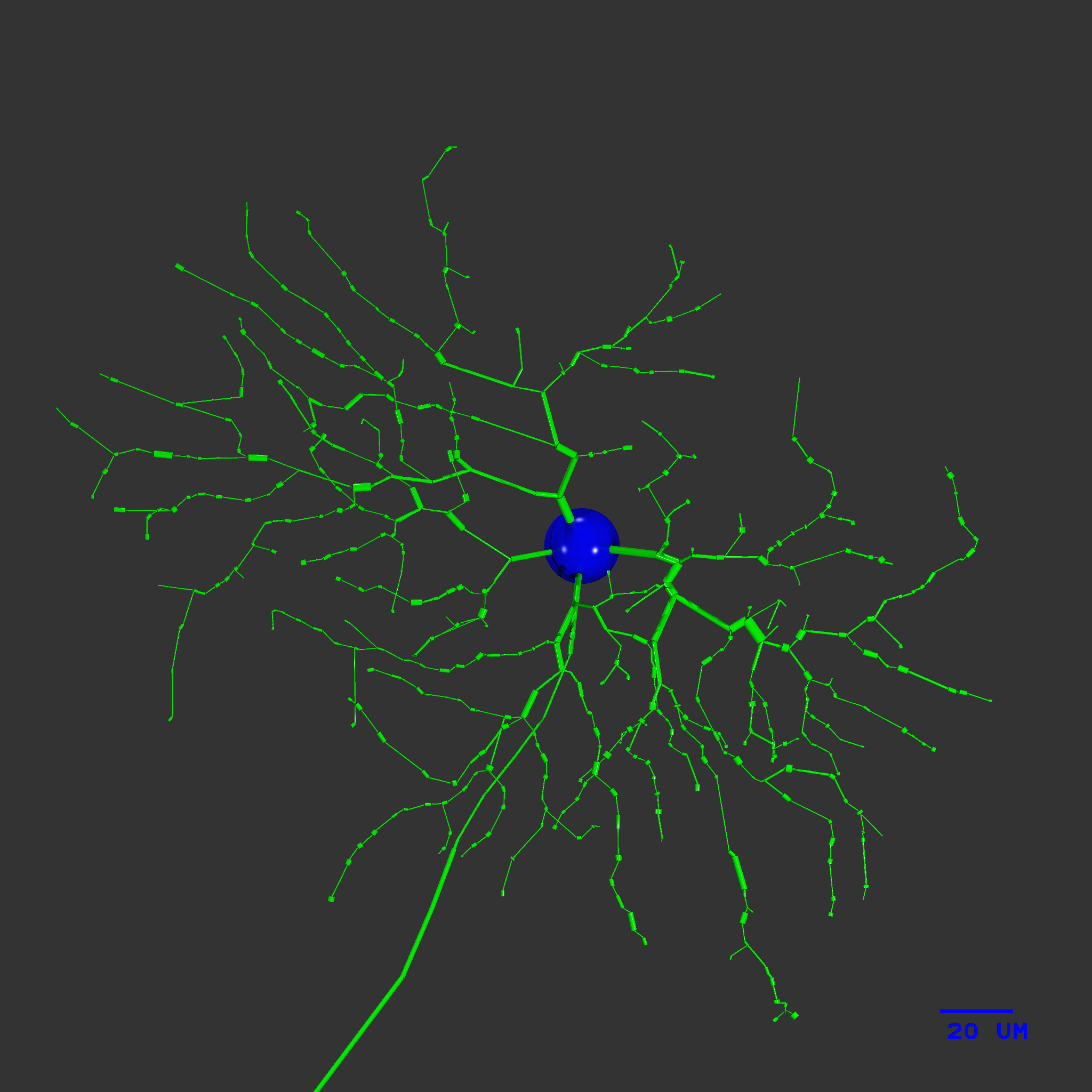
Model of small central (1 degree ecc.) on-beta (X) ganglion cell showing 20 um dia soma, dendritic tree, axon hillock and axon initial segment.
We are interested in understanding what limits the signal transmitted by a neuron. A spiking neuron collects signals from synaptic inputs in its dendritic tree. A sequence of action potentials, or “spikes” is generated by a collection of membrane ion channels, and the spikes are transmitted along the neuron's axon. This conversion from analog signals to all-or-nothing spikes allows the neuron to pass information to other neurons, but it also provides an opportunity for signal processing, either to modify the signal's amplitude, change its temporal properties, or vary them according to the input signals. How this process is accomplished is an important question in Neuroscience.
The ganglion cell receives synaptic contacts from an array of several hundred bipolar cells spaced regularly throughout its dendritic tree. Each synapse transmits its signal using vesicles of glutamate, called EPSPs or "quanta" by physiologists who record the synaptic responses. The vesicles are released to some extent randomly by the bipolar cell, but the rate of release is modulated by the bipolar's presynaptic voltage. Therefore the signal in the ganglion cell is noisy, which limits the precision of the ganglion cell's signal. However, the ganglion cell spike generator contains voltage-gated channels, which are also noisy.
We have studied how the ganglion cell's intrinsic noise sources affect its signal with a computational model (van Rossum et al., 2003). We have also studied this effect by recording ganglion cell responses to a flash of light, and comparing the variability of the generator potential and spike train (Dhingra and Smith, 2004). From these studies it is apparent that the ganglion cell spike generator loses about half the gray levels available to it from synaptic input.
Voltage recording from soma with different rates vesicle release applied a synaptic inputs. Bottom trace: record of vesicle release events at one synapse, scale: fraction of synaptic channels bound with neurotransmitter, saturating at 1 (center ordinate). Middle trace: rate of vesicle release at one synapse, scale: events/sec (leftmost ordinate). Top trace: record of voltage noise in ganglion cell showing linearity of response and noise amplitude at different rates of input, scale mV.
- Rob